Skin tumors are rather common. According to the data available in the literature, malignant neoplasia of the skin affects 3 to 75 per 100,000 individuals in different countries of the world. The incidence of pretumors and benign diseases of the skin is 3-4 times higher than that of malignant ones (I.R.Lazarev, 1973).
According to their clinical presentation, neoplasms of the skin are divided into malignant, benign, and precancerous ones.
There are several treatments for skin tumors. These include radiation therapy, chemotherapy, surgery, and multimodality treatment. None of the currently available treatments can completely answer the surgeon's requirements so there is a need to search for new solutions of the above problem.
In this connection, the use carbon-dioxide laser is of great interest. It should be noted that the impact of laser radiation on biological structures is specific and different from all other known physical expo-sures. The basis for the interaction of laser radiation with the skin and subcutaneous fat is the physical properties of laser radiation itself and the physicobiological properties of the skin and adjacent tissues. The physical factors of laser radiation include the rate of acting luminous energy, the wavelength of light, its monochromatism, polarization, coherence, radiating conditions, and exposure time. The physical and biological properties of the skin involve its reflecting and absorbing capacities, heat conduction and capacity, the poten-tial of the skin, the degree of its pigmentation and vascularization. The use of carbon-dioxide lasers in skin plastic repairs to treat cutaneous tumors is justifiable and promising. Such advantages of the laser over other cutting instruments as adequate hemostasis, sterility of an area to be operated on, and minimum tissue traumatization permit its use during various surgical interventions.
Depending on the spread and site of benign tumors, the following surgical interventions are indicated:
1) laser photocoagulation;
2) laser excision of a tumor along with stitching of the wound margins;
3) laser excision of a tumor along with plastic repair with local tissues.
The indications for this treatment are tumors, under 1 cm in diameter, if they are on the face and neck, and those, no more than 2.5 cm in diameter, in other parts of the body. Laser photocoagulation is also indicated if benign tumors are present on the inferior or superior margin of the wings of the nose, on its tip, in the cutaneous part of the nasal opening, on the inner surface of the concha auriculae, in the upper and lower eyelids, in the anal canal or the areas inaccessible for scalpel surgery, and when operations using a scalpel may cause organ deformity or yield a poor cosmetic result.
The procedure of laser photocoagulation is as follows. The tumor itself and adjacent tissues are maximally infiltrated with 0.25-0.5% trimecaine or novocaine solution. This is necessary not only for anesthesia, but for creation of a protective liquid barrier keeping the surrounding tissues from the temperature action of a laser beam. Tumor vaporization is generally started with the periphery, with the boundary of healthy tissue by gripping as large as 1-mm normal tissue rim and a laser beam is directed to the center of the tumor. It is expedient to make laser photocoagulation with a continuous focused laser beam 0.5 mm in diameter, at 3-5 W, with a power density of 0.25-1.0x10³ W/cm2. The laser radiation power varies with the site of a tumor as the thickness of the skin in different areas of the body is variable.
Table 7 gives the thickness of the skin, epidermis, and der-mis in man in different body's parts which allow the surgeon to simulate the parameters of laser radiation.
It is undesirable to increase the power up to greater than 10 W on vaporization as with this it is more difficult to control the depth of laser beam penetration and the formation of a coagulation necrotic zone. Its increase may exert an adverse effect on beauty treatment outcomes. To prevent recurrence, photocoagulation of benign tumors should be made until the carbonized tumor parts resulting from the burning of dense connective tumor tissue parts, which may extend into the subcutaneous fat, disappear in the wound. After surgery, the wound is cleansed with 5% potassium permanganate solution. A bandage should not be applied.
Table 7
| Skin region Dermis | Epidermis | Dermis | The whole skin |
|---|---|---|---|
| Head | |||
| forehead | 0.1 | 2.1 | 2.2 |
| hairy part | 0.2 | 2.4 | 2.6 |
| nose | 0.1 | 2.1 | 2.2 |
| upper lip | 0.2 | 1.8 | 2.0 |
| Chest | 0.1 | 3.0 | 3.1 |
| Abdomen | 0.1 | 2.3 | 2.4 |
| Pubic region | 0.1 | 1.7 | 1.8 |
| Back | 0.1 | 4.8 | 4.9 |
| Gluteal region | 0.2 | 3.0 | 3.2 |
| Arm | |||
| anterior surface | 0.1 | 2.1 | 2.2 |
| posterior surface | 0.1 | 3.0 | 3.1 |
| Forearm | |||
| anterior surface | 0.1 | 1.8 | 1.9 |
| Leg | |||
| anterior surface | 0.1 | 2.3 | 2.4 |
| posterior surface | 0.2 | 3.1 | 3.3 |
| Hand | |||
| back | 0.3 | 2.7 | 3.0 |
| palm | 0.7 | 1.9 | 2.6 |
| Foot | |||
| back | 0.3 | 2.0 | 2.3 |
| sole | 1.0 | 1.7 | 2.7 |
The indications for this surgical treatment are benign tumors, above 1 cm in diameter, if they are on the face and those, no more than 2.5 cm in diameter, in other parts of the human body. This group mainly comprises fibromas, lipomas and papillomas attached by a broad base, precancer of the skin, retention cysts of the mucosa, and atheromas. These surgical interventions are chiefly made under local anesthesia by 0.25-1.0% trimecaine or novocaine solution.
Before all operations, the borders of removed tissues should be marked on the skin with brilliant green. This considerably promotes both the planning of the operation itself and its net result.
After incising the dermis with a scalpel, the removed part with an abnormal focus is taken on the holders and the tumor is excised using a pulsed focused laser beam, the diameter of a light spot is 0.5 mm, the power 5-7 W, the duration of a pulse 0.05 sec, the pulse pause 0.1 sec, the energy power will be 7-8 J/mm2 (see Tables 5 and 6). When the tumor is separated with the laser beam, tiny blood vessels are sealed, the hemostatic clamps should be applied to the vessels above 1 mm in diameter. After laser dissection of these vessels, the clamp is removed without applying catgut ligatures since the walls of bloodless vessels are sealed between themselves in nearly all cases. The wound is closed with interrupted sutures using an atraumatic needle and a synthetic monophilic thread (4/0-6/0). Sutures in the skin and on the face are usually removed on days 7-8 and 3-5, respectively.
Operations with plastic repair with local tissues are indicated in patients with large benign tumors, pigmented warty nevi, fibrolipomas, or with tumors in the body's parts, such as the forehead, the hairy part of the head, parts, the anterior surface of the leg, wherein the operation using the closure of the wound with sutures directed towards the surgeon is impossible to perform for this or that reasons. These tumors average 3-5 mm or more in diameter.
If the surgical intervention is bulky, extensive, and traumatic, it is performed under intravenous or general intubation anesthesia. Here, too, indifferent solutions (0.25-0.5% trimecaine or novocaine solution) should be, however, administered into the area under operation to protect the surrounding tissues from the temperature action of a laser beam.
The skin is dissected to its basal layer with a routine scalpel along the premarked line around the tumor. The latter is fixed with Kocher's clamps or holders, then separated with a focused laser beam. With the pulsed operation, the radiation power is 5-7 W, the duration of a pulse 0.05-0.1 sec, the pulse pause 0.1 sec. Laser tissue separation is started with the periphery. On entering the fluid layer, the power may be build up to 15 W, showing an increase in the rate of tissue separation. The risk of adjacent tissue injury is virtually ruled out due to the protective layer of the indifferent solution injected. The procedure of hemostasis is the same as that during the operations described above. After cutting out a skin graft for plastic repair, the wound at the donor site is closed in layers, by plunging catgut sutures with knots inwards, those to the skin using an atraumatic needle and synthetic monophilic thread (4/0-6/0). The cutaneous fat graft is fixed to the defect margins with interrupted sutures (5/0-6/0) by using only a synthetic monophilic thread and an atraumatic needle. The sutures are removed on days 7-8 after surgery.
In pedunculated tumors (fibromas, papillomas) and in tumors of the eyelids, tendinous and articular ganglia, they may be dissected using the laser clamps. After giving local infiltration anesthesia by 1% novocaine solution, a laser clamp is, at a distance of 1 cm from the tumor base, applied to the tumor peduncularly to the base by gripping the adjacent skin parts. The continuous focused laser beam with a 0.2-mm spot at an output power of 10 W (the power density is 32x103 W/cm2) is used to cut off the tumor so that the protruding part of the base with the adjacent skin is left in the clamp. The tissue remainder is removed via vaporization which is made by a defocused laser beam, 0.7 mm in diameter, the spots of pilot radiation being set apart up to 1 cm (the power density is 2.6x10³ W/cm2). When the instrument is removed, at the site of the tumor there is a rather solid linear coagulation crust whose fixation does not generally require suture application.
The anatomic peculiarity of the eyelid skin (extreme thinning and high mobility) makes it possible to use this method even in the presence of sessile tumors. After injection of 1% novocaine solution beneath the eyelid skin, the latter assembles as a fold in the center of which the tumor is located. The laser clamp is applied to this fold. Due to the close proximity of the eye-ball, the neighbouring tissues are protected with saline-wetted drapes. The further procedures of the operation are the same as described above.
On removing the tendinous ganglia, a clamp is applied transversely to the run of the tendon in order to prevent its deformity. On removing the articular ganglia, the direction of applying the clamp is not of fundamental importance. The formed coagulation crust hermetically seals the ganglionary base. In the postoperative period, immobilization should be made within a week.
To treat patients with malignant tumors of the skin presents a real challenge and depends on the timely detection of the disease at stages I and II. Surgical interventions using the routine scalpel are traumatic, which may promote penetration of cancer cells into the lymphatic and blood channels and metastatic spreading from the basic focus.
The surgical laser is an aid in circumventing these difficulties and making not only radical and palliative tumor removal, but also, if required, skin plastic repair, i.e. cosmetic intervention. The efficiency of surgical treatment in cancer patients enhances due to the sealing of blood and lymphatic vessels via laser radiation.
The indications for laser radiation are basal and squamous cell carcinomas of the skin, melanoma, disseminated melanomas, and precancers of the skin and mucosa. There are virtually no contraindications to the use of laser radiation in the surgical treatment of malignant tumors.
Laser radiation is successfully employed in the treatment of recurrent and inoperable cutaneous and mucosal tumors complicated by hemorrhage, ulcerations, superinfection, and pain. Despite the fact that these complications are not generally life-threatening when the patient visits a doctor; nevertheless, they make his/her social adaptation difficult. It is very significant that excision and/or treatment of this tumor with a defocused carbon-dioxide laser beam relieve the patients of pain, obnoxious odour, inferiority, and diffidence.
To treat malignant tumors of the skin, the following surgical interventions are indicated:
1) laser photocoagulation;
2) laser excision of a tumor along with stitching of the wound margins;
3) laser excision of a tumor along with plastic repair with local tissues;
4) laser excision of a tumor along with plastic repair with free skin grafts.
This procedure is indicated in patients with T1N0M0 basal and squamous cell carcinomas of the skin and mouth, whose size is no more than 0.5 cm in diameter. The surgical techniques are as follows. The skin and subcutaneous fat at the site of a tumor are infiltrated with 5-10 ml of 0.5% novocaine solution. At a distance of 3-4 cm from the visible border of the tumor, the skin with the abnormal formation, from periphery to center, is circularly vaporized its whole dermal thickness to the subcutaneous fat layer, by using a laser beam. The radiation mode is continu-ous, the power is 10-15 W, the diameter of a focused laser beam 0.2 mm spot. The power density is 5.1-7.6x10³ W/cm2). The laser radiation power is sufficient to completely vaporize the pathological formation for 40-60 sec without ther-mally damaging the surrounding tissues. This shows a good hemostasis, ablasticity and sterility of the wound surface. The wound is cleansed with 5% potassium permanganate solution, a bandage is not applied. Insignificant edema persists within the first 2 postoperative days. The patients have virtually no complaints of pain therefore no analgetic therapy is generally given.
This procedure is indicated in patients with T2N0M0 basal and squamous cell carcinomas of the skin and mucosa, whose size ranges from 0.6 to 2.5 cm in diameter. The surgical techniques are as follows. The skin and mucosal membrane are incised with a routine scalpel at a distance of 6-8 and 15- 18 cm from the visible border of the tumor in the presence of basal- and squamous-cell carcinomas, respectively. 0.25-0.5% novocaine or trimecaine solution is preadministered beneath the tumor. The pulsed 0.5-mm focused laser beam at 5-7 W (the pulse duration is 0.05-0.1 sec, pause is 0.1 sec) is used to excise the tumor in the marked limits. Our investigations have demonstrated that the incision of the skin with the focused laser beam after its infiltration with indifferent solution is followed by the formation of 100-150-µm coagulation necrotic area. The tissues are separated in the strictly predetermined layer. The laser-induced wound usually heals in the same period as does the scalpel-induced one; however, the laser yields a better cosmetic effect. On the completion of the operation, interrupted sutures are applied using the synthetic monophilic thread (4/0-6/0) in the atraumatic needle.
This surgical treatment is indicated in patients with malignant tumors (T1 and T2) of the skin in the presence of palpable regional lymph nodes and in the absence of distal metastases, in those with tumors of the maxillofacial region, hand, or tumors (T3N1M0) of other parts of the body. The diagnosis should be verified by paracentetic and cytological or morphological studies from the data of biopsy.
The surgical techniques and procedure are as follows. The lines and borders of excised tissues and graft separation are premarked with brilliant green. 0.5-1.0% anesthetic solution is administered beneath the tumor and adjacent tissues. Then the skin is dissected along the marked line to the basal layer using a scalpel and further tissue separation is performed with a 0.5-mm focused carbon-dioxide laser beam at 7-10 W. The radiation mode is pulsed, the pulse duration is 0.05-0.1 sec, the pause 0.1 sec). This procedure for laser tissue dissection has been developed under the supervision of Professor O.K.Skobelkin at the State Research Center of Laser Medicine, Ministry of Health and Medical Industry, and named laser photohydraulic tissue dissection (USSR Author's Certificate No. 628647).
Local plastic repairs generally end with the fixation of grafts with interrupted sutures and the stitching of the flap margins to the bed using the 4/0-6/0 thread and atraumatic needle. Bandages are not applied after operations on the head and neck. The sutures are removed on postoperative days 7 to 9.
This surgical procedure is used in patients with squamous- and basal-cell skin carcinomas located in the forehead, temporal region, hairy part of the head, palms, talocriral articulations, i.e. in the areas wherein it is impossible to close a wound defect with tissues or to suture. It is a very good matter to use laser photohydraulic dissection. Early at surgery, when the borders of a graft and tumor excision are marked, it is advisable to apply a 0.5-mm focused laser beam. The radiation mode is continuous, the power is 5-7 W. On mobilizing the tumor, the radiation power may be increased up to 10-15 W.
The formed free skin graft is stitched to the defect margins with interrupted sutures using a 4/0-6/0 synthetic monophilic thread and an atraumatic needle. The wound at the donor site is sutured with a 4/0 monophilic thread. If the wound cannot be closed with sutures "directed towards the surgeon"), plastic techniques (mobilization of the wound margins, additional incisions, back triangular grafts) are employed. During operations on the head and neck, grafts for free plastic repair are taken from the parotic region or the inner surfaces of the arm. While making operation on other parts of the body, free skin grafts are taken from the inguinal regions of the abdomen, the lateral regions of the chest, and the inner surfaces of the legs.
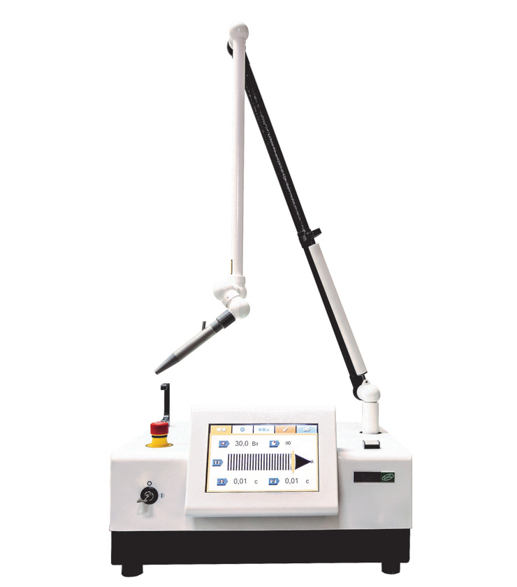
The use of laser photohydraulic dissection during skin plastic operations substantially decreases the adverse effects of laser radiation on skin grafts and surrounding tissues, by reducing the thermal action of laser radiation on tissues to a minimum. The used procedure for tissue dissection creates favourable conditions for wound healing in the postoperative period. Peculiar laser coagulation thrombi are formed in the vessels due to their local thermal effects, through which tissue dissection is bloodless and the coagulation of nerve endings causes no terrible ache. Laser radiation provides operative field sterility, substantially reducing the incidence of suppurations and graft rejections. Wounds heal in the usual periods of time to give rise to delicate elastic cicatrices and to yield good cosmetic and functional results. The proposed procedures using the Lancet LSA in the surgical treatment of tumors depending on their size, site, stage, and the power of laser radiation allow the practitioners to choose a procedure of laser surgical treatment correctly and to achieve good late outcomes.
The Lancet LSA may be successfully used in plastic repairs in surgical cosmetology, such as reduction mammoplasty, abdominoplasty, blepharoplasty, otoplasty, and lifting. Due to its physical characteristics, a surgical laser fits as a scalpel to dissect varying density tissues. The Lantset LSA provides sensory change-over and is able to decrease or increase the power of and change other parameters of laser radiation, which allows the laser operating mode to be changed intraoperatively. This is very convenient to operate on varying density structures with good hemostasis and operative field sterility.
Reduction mammoplasty is an operation to correct the breast. It consists in removing excess skin and stroma of the gland itself. Different laser radiation powers are used depending on the surgical stage. The continuous laser beam, 0.5 mm in diameter, at as high as 0.5 W (power density is 2.5x10³ W/cm2) is used to make out on the operating table. This enables the skin marks to be kept till the end of the operation. Pulsed laser radiation (pulse duration 0.5 sec, power 10 W) is used at the stage of graft adermatization. The continuous laser beam, focused to 0.5 mm, at 15 W is applied when resecting the stroma of the gland itself.
The use of a Lancet in reduction mammoplasty permits its rapid performance with good hemostasis in aseptic conditions, i.e. all necessary conditions under which the optimum result is achieved.
Abdominoplasty is mainly the correction of the skin, subcutaneous fat, muscles and fasciae of the anterior abdominal wall for cosmetic purposes. It is reasonable to use a carbon-dioxide laser at the stage of separating skin fat grafts. The skin is dissected to the subcutaneous fat with a scalpel and further separation is made using a continuous laser beam focused to 0.5 mm at the power of as high as 20 W. The muscles and aponeurosis are sutured with a 2/0 lavsan (dacron) thread. The skin is stitched with an intra-dermal suture using a 4/0 prolenic one.
Lifting is a cosmetic operation for removing excess skin on the face. During this operation, a focused laser beam is used in combination with photohydraulic dissection, which prevents its thermal effects on the surrounding tissues and the possible traumatization of the adjacent parts (fasciae, branches of the facial nerve and temporal vessels). A continuous laser radiation (at 10 W with a spot diameter of 0.5 mm) is applied to separate a skin graft.
Blepharoplasty is an operation for eliminating excess skin on the lower and upper eyelids and for removing periorbital hernial sacs. During this plastic operation, laser photohydraulic dissection is also made to remove excess skin. The laser beam is focused to 0.3 mm, the mode is pulsed, power 5 W, the duration of a pulse 0.05-0.1 sec. The operation is quite bloodless and prompt. The skin is sutured using a 6/0 vicryl thread.
Otoplasty is a plastic operation to correct the concha auriculae. This operation requires the use of pulsed radiation at 5 W (pulse duration 0.05-0.1 sec, pause 0.1 sec,) the beam is focused to 0.5 mm. The same parameters of laser radiation are employed for vascular coagulation. The skin is stitched with a continuous catgut suture.
Представительство в Москве


Представительство в Туле